Service
1 Technical consultation and services
1.1 Contact
Tel: 0577-88017533
Email: support@sensby.com
QQ: 3289287609
1.2 Technical service content
- Different biomolecular interaction tests (protein protein, antibody antigen, drug cell, DNA-DNA, polypeptide cell, gas chemical molecule, etc.) and give the intermolecular affinity and dissociation constant
- Film thickness and refractive index test (including biofilm, solid film)
- Drug delivery and drug release testing
- Antibody screening, drug screening
- Application test of biomaterials and tissue engineering
- Detection of biosensors
- Electrochemical testing
2 Application cases
Case 1
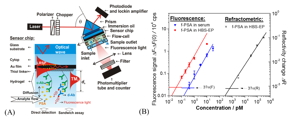
Using SPR sensing technology, we can detect prostate specific antigen (PSA) in serum. We modified the surface of SPR chip with carboxymethyl dextran (PCMD) and used it as a 3D binding matrix with non-specific adsorption ability to detect PSA in serum (Fig. 1A). The detection sensitivity of this method can reach 34fM (Fig. 1B).

Figure 1 (A) schematic diagram of SPR enhanced fluorescence device and surface modification of PCMD on SPR chip as binding matrix of antibody molecules, (B) standard curve of direct detection of PSA by SPR enhanced fluorescence and SPR label free in buffer and serum. (quoted from: Wang Y.#, et al., Anal. Chem. 2009, 81, 9625–9632)
Case 2
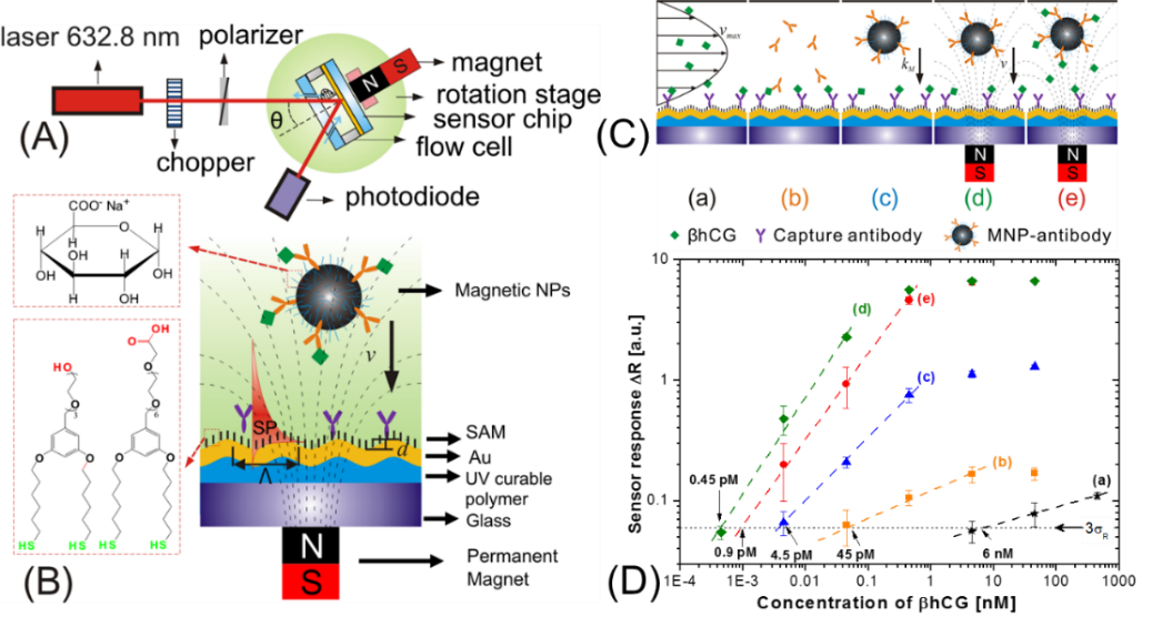
Using SPR technology combined with magnetic nanoparticles, the detection of human chorionic gonadotropin (hcG) in 15 min was achieved, and the sensitivity was 0.45 pM. In this method, we use magnetic nanoparticles as carriers to transport target molecules to the surface of SPR sensor chip under the action of magnetic field, and amplify SPR signal. We used a grating coupled SPR method and applied a magnetic field on the back of the chip to control the magnetic nanoparticles in the sample solution (Fig. 2A, B). The iron oxide magnetic nanoparticles and metal grating chips used in this method are modified with specific antibodies to form sandwich structure detection model. At the same time, we studied the relationship between the sensitivity of biosensor and molecular diffusion or magnetic field gradient (Fig. 2C). Finally, we used immunoassay to detect hcG with high sensitivity. Compared with the traditional SPR detection method, the sensitivity is improved by 4 orders of magnitude, reaching the detection limit of 0.45pM (Fig. 2D), and the detection is completed within 15 minutes.

Figure 2 (A) grating SPR device, (B) grating chip combined with magnetic nanoparticles and magnets to accelerate the adhesion of the tested substance to the gold surface, (C) different modes of SPR sensors, including (a) direct label free hCG, (b) antibody amplification signal, (c) magnetic nanoparticles enhancing SPR, (d) accelerating the binding of magnetic nanoparticles on gold sheet under the action of magnets, and (e) integrating magnetic nanoparticles into gold wafer The magnetic nanoparticles are pre bonded to hcG, and the magnetic field accelerates the binding of the tested substance to the gold sheet. (quoted from: Wang Y.#, et al. Anal. Chem., 2011, 83, 6202-6207.)
Case 3
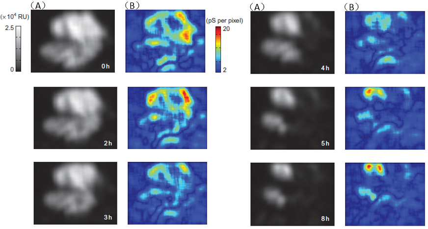
We can study the dynamic process of single cell (such as apoptosis, electroporation, etc.) by using high-resolution microscopic imaging built by SPR technology.
In Figure 3, SPR high-resolution microscopic imaging is used to detect the interaction between drug molecules and virus cells. As shown in the figure, the apoptosis process of SiHa cells after the addition of MG132 and TRAIL. It can be clearly seen from Fig. 3 (B) that after MG132 and TRAIL enter SiHa cells, the nuclear materials of SiHa cells begin to divide, resulting in the whole cells starting to contract, and then some bubbles are formed near the cytoplasmic membrane and finally disintegrated.
 Fig. 3(A) SPR of SiHa cells and (B) EIS image (quoted from: Wang, W.; Tao, N et al. Nature Chemistry 2011, 3, 249)


